

Article: Microscopic Views of Chrysocolla

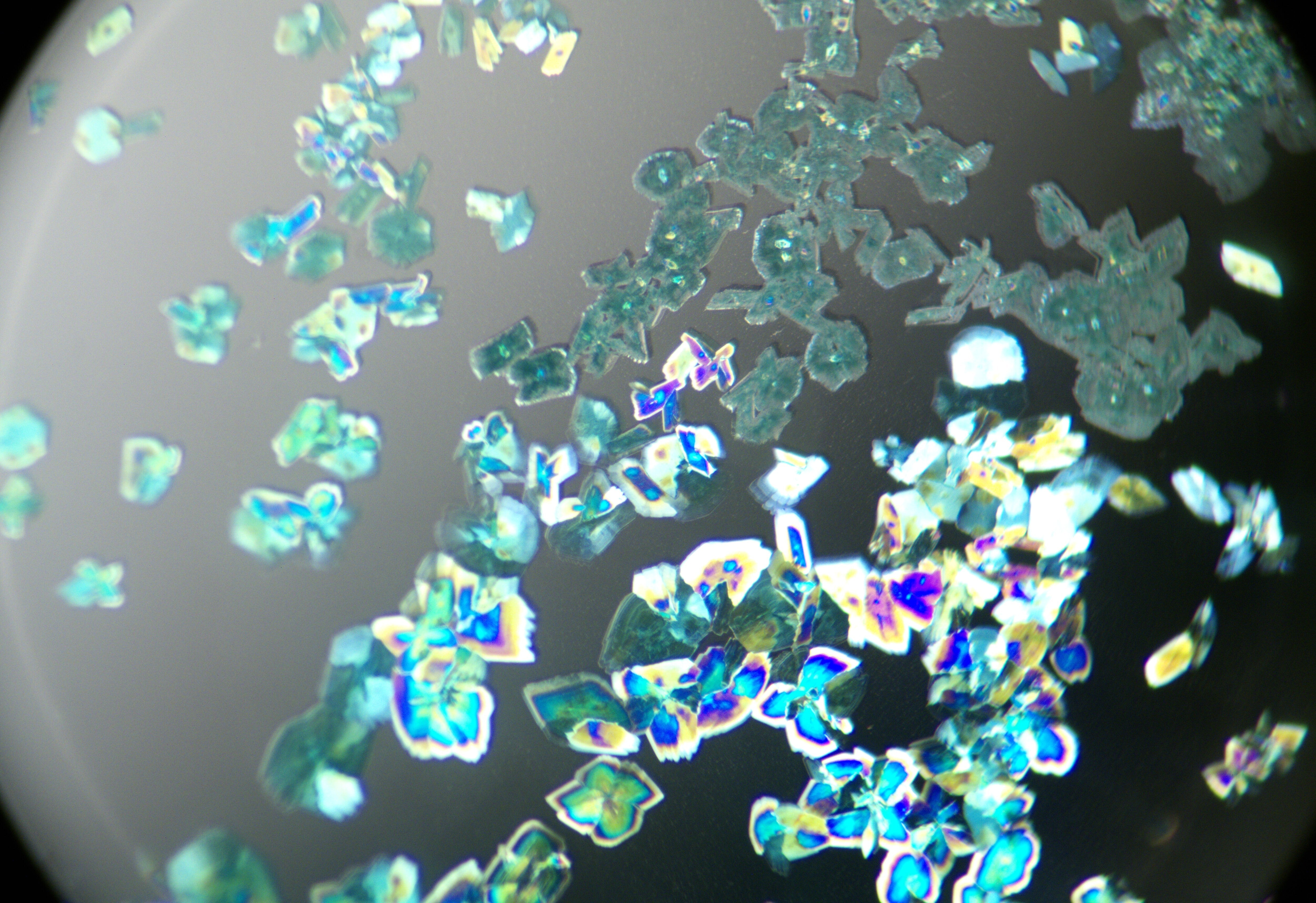
Microscopic Views of Chrysocolla

Figure 1 — Mineral crystals at 15× magnification. Rhombic and pseudohexagonal platy forms are visible, together with a bull's-eye interference figure.

Figure 2 — At 20× magnification under crossed polars, iridescent interference colours are visible across the crystal surfaces.

Figure 3 — A broader view of the mineral crystals at 12× magnification.

This chrysocolla specimen is described by the supplier as an uncommon example from a small group of six. It is the only visibly clean specimen in that group. The material is bicoloured: one half is opaque pale blue-green, while the other is transparent deep blue-green. It has perceptible thickness and displays thin, rhombic to pseudohexagonal platy crystals. Earlier examples seen by the supplier reportedly contained more abundant cloudy areas and fissures.


